
A laser needle to observe microscopic objects
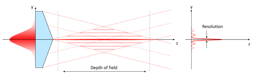
Bessel beams are “needles” of light formed by the interference of a laser beam with a conical-phase (Fig. 1). They have many applications in both research and industry. In particular, they are used for machining transparent materials, such as glass or sapphire, and to make smartphone screens and watch glasses. Their properties also make them particularly attractive for viewing incredibly small structures and observing biological phenomena that are invisible to the naked eye. Why are Bessel beams useful for microscopy?
Laser microscopy techniques
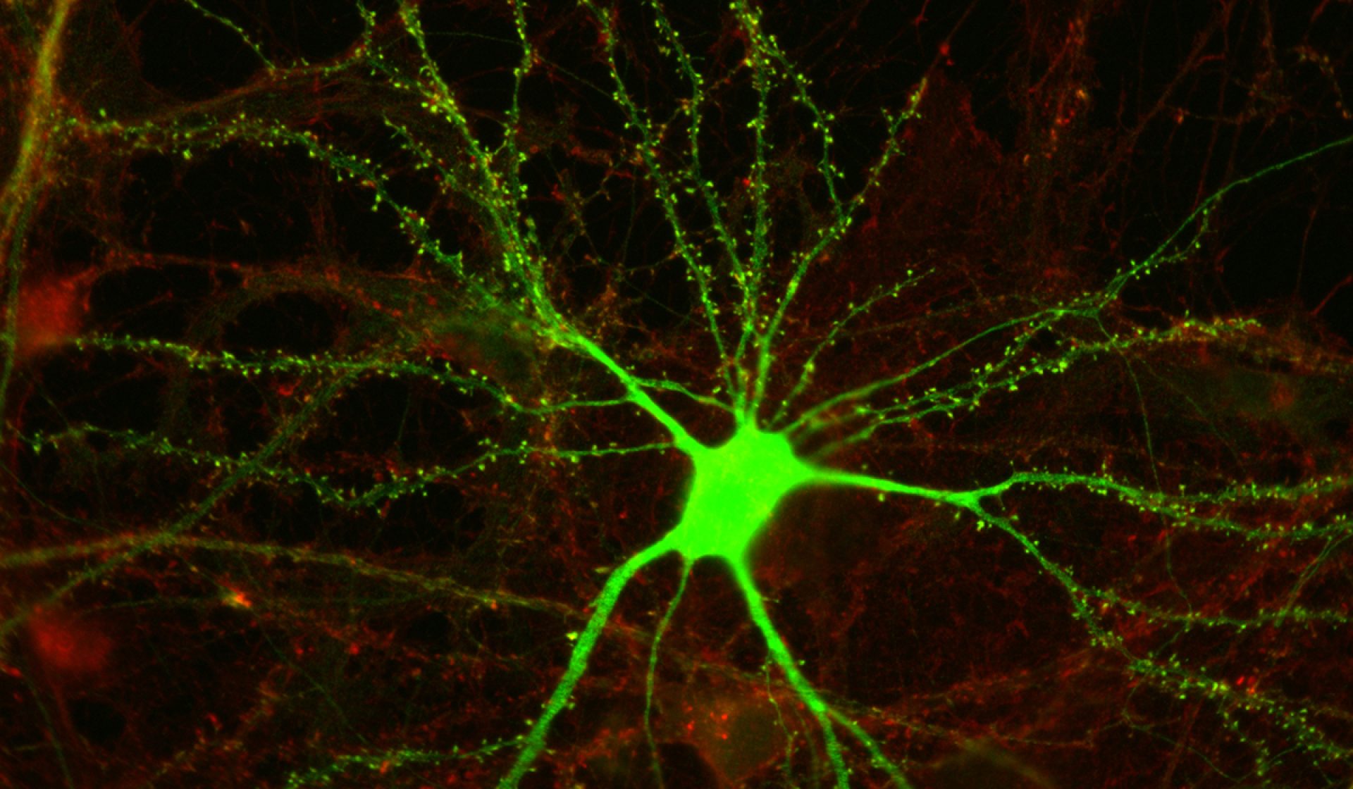
The various laser microscopy techniques, such as light-sheet microscopy or fluorescence microscopy, are indispensable tools for biologists. For example, two-photon microscopy, a type of fluorescence microscopy, allows the imaging of biological structures in tissues and can even use fluorescent tracers to monitor the activity(1) of cell networks. It is therefore a particularly useful technique for neuroscience (interesting fact: the two-photon absorption theory was developed in the thesis of the brilliant Maria Goeppert Mayer, who went on to win the Nobel Prize in Physics).
These techniques are all based on a laser beam. However, the conventional (Gaussian) focusing of a laser beam is not ideal and is even limited in some applications. Bessel beams have properties that are particularly suitable for microscopy and are increasingly used in new applications. They have been used for several years to improve laser microscopy techniques. So what are the benefits of Bessel beams for microscopy?
- Depth of field
A conventional Gaussian beam only offers a shallow depth of field. This can be useful because it allows us to image a sample plane by plane. However, this technique is too slow when it comes to imaging larger volumes such as the network of neurons that makes up our brain. In addition, if the observed object is alive (in vivo observation), small movements such as breathing can distort the measurements(1).
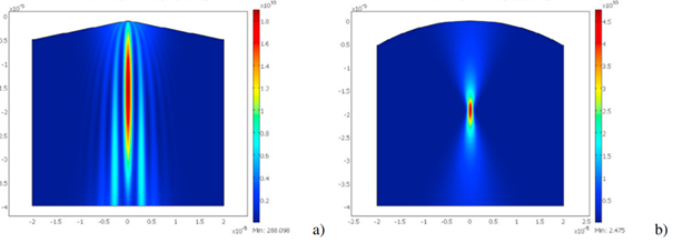
A Bessel beam has a depth of focus that is 10 to 100 times longer than a conventional focus(2) (Fig. 2). By using a Bessel beam to illuminate and image a sample, researchers can considerably increase the depth of field, making it possible to image a volume much faster, and thus observe live events taking place in a volume while resisting any small movements of the object being observed(3).
- Spatial resolution
It is important to be able to image a biological sample with a resolution smaller than the size of the living cells being studied. For example, a resolution of a few hundred nanometers is required(1) to observe cell structures. One of the advantages of Bessel beams is that they are not limited by the diffraction limit: the dimensions of their focus can be smaller than the wavelength used, so Bessel beams can have a width of the order of a hundred nanometers, providing excellent imaging resolution.
- Damage to samples
Observing biological tissues using a laser microscopy technique can disturb or even damage sensitive samples if they absorb the light energy emitted. Because of the extreme depth of focus of Bessel beams, the light energy is more “spread out” than for Gaussian beams. This allows samples to be imaged by exposing them to lower light intensity, thereby reducing any potential for damage(1).
- Resistance to obstacles and disturbances
A Bessel beam is generated along the propagation axis by the interference of photons arriving from the side (Fig. 1). This gives it the property of being “regenerative”, i.e. the beam rebuilds itself if it encounters an obstacle along its propagation(4). This property is especially used for selective plane illumination microscopy: this technique uses a laser beam to illuminate a sample from the side and if the sample is too thick, the laser beam scatters light, reducing the image quality. The regenerative property of Bessel beams means that the image quality is enhanced, even for thick samples. Researchers at the University of Freiburg (Germany) have applied Bessel beams to imaging in order to visualize 50% deeper inside human skin than with a conventional beam(4).
CANUNDA-AXICON
Cailabs has been developing fully reflective axicons for high precision applications since 2017. The manufacturing precision of these axicons can generate Bessel beams with exceptional quality, while avoiding the chromatic aberrations and energy oscillations typical of conventional refractive axicons. This quality makes CANUNDA-AXICON the perfect tool for imaging and microscopy applications.
Sources:
(1) Extended two-photon microscopy in live samples with Bessel beams: Steadier focus, faster volume scans, and simpler stereoscopic imaging (2014), Gabrielle Thériault et al.
(2) Application of Bessel beams for ultrafast laser volume structuring of non-transparent media (2010), Alexeev et al.
(3) Rapid volumetric imaging with Bessel-Beam three-photon microscopy (2018), Bingying Chen et al.
(4) Bessel beam allows microscope to look deep into tissue (2010), John Wallace

By Sami Laroui
Sami Laroui, who holds Master’s degrees in materials science and innovation and entrepreneurship, joined Cailabs in 2018. As a pre-sales engineer, he contributes to the development and commercialization of innovative optical solutions, that optimize the quality and performance of laser machining processes.
Our Other Articles:
-

Exceeding Throughput Limits with Laser Communications
-

The challenges of e-mobility: Welding busbars
-

Flying further with beam shaping
-

From the Volkswagen Golf to the Tesla Model 3: How Lasers Are Shaping the Automotive Industry?
-
Space Optical Communications: Why Are Space-to-ground Links Taking Time to Develop?